Morphology of vestibular aqueduct and endolymphatic sac
Barbara A. Bohne, Ph.D. Professor of Otolaryngology and Valentin Militchin, M.S.
ANATOMY of the HUMAN TEMPORAL BONE
HORIZONTAL SERIES of SECTIONS through HUMAN TEMPORAL BONE
本系列组织切片显示了前庭水管及内淋巴囊与总脚、后半规管的组织和解剖关系。
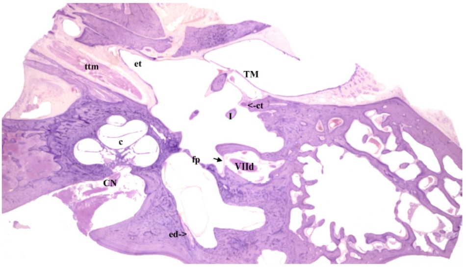
Section 181
The origin of the endolymphatic duct (ed) is visible at the origin of the vestibular aqueduct.
c – Cochlea; CN – cochlear division of VIIIth CN; ct – chorda tympani; et – Eustachian tube; I – long process of incus; TM – tympanic membrane.
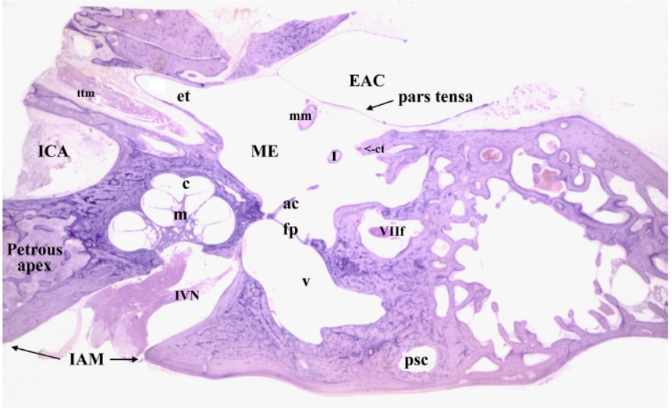
section 200
The tympanic membrane (eardrum) forms the boundary between the external auditory canal (EAC) and middle ear (ME). The manubrium (mm) of the malleus is attached to the medial surface of the eardrum. The long process of the incus (I) is located medial to the manubrium and lateral to the stapes (ac – anterior crus). Anteriorly, the middle ear narrows down to form the bony portion of the Eustachian (auditory) tube (et) which connects the middle ear to the nasopharynx. Running parallel to the auditory tube is the tensor tympani muscle (ttm) in its bony semicanal. The chorda tympani (ct) is nearing the posterior wall of the middle ear space. The footplate (fp) of the stapes which is held in the oval window by the annular ligament forms the boundary between the middle and inner ears. In this section both the cochlear and inferior vestibular portion (IVN) of VIIIth CN are visible. Note that the cochlea is anterior to the vestibular portion of the membranous labyrinth. In the cochlea, 5 cuts through the coiled cochlear duct are seen as it spirals around the modiolus (m). The vestibule (v) and the posterior semicircular canal (psc) are visible. IAM – internal auditory meatus; ICA – Internal carotid artery; VIIf – Mastoid segment of facial nerve.
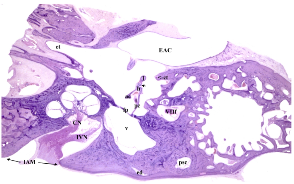
section 221
Nearly the complete stapes is present: ac – anterior crus; fp – footplate; h – head; pc – posterior crus. The incudostapedial joint between stapes head and lenticular process of the incus is visible. The lenticular process (arrow) is connected to the long process (I) of the incus by a connecting stem (Grayboyes et al., 2011). The chorda tympani (ct) has reached the posterior wall of the middle ear space at the posterior iter. Both the cochlea and vestibule (v) are still present. Only the non-sensory portion of the posterior semicircular canal (psc) is visible. The endolymphatic duct (ed) can be seen within the osseous vestibular aqueduct. The inferior division of the VIII nerve with branches to the cochlea (CN) and saccule (IVN) is visible in the internal auditory canal. EAC – external auditory canal; IAM – internal auditory meatus; VIIf – Mastoid segment of facial nerve.
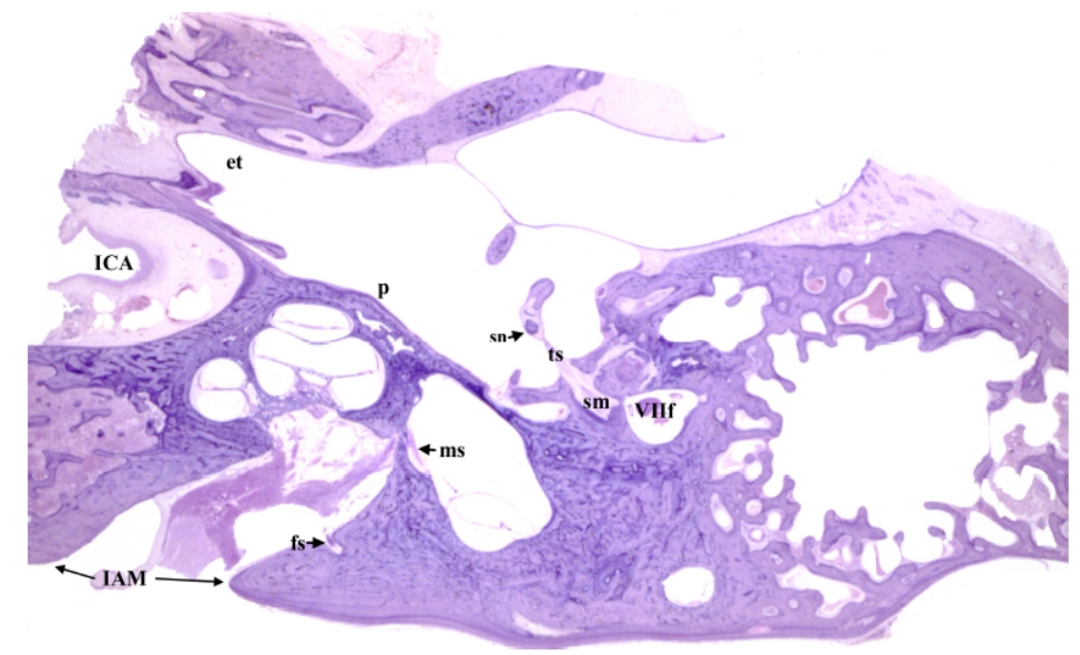
section 241
The stapedius muscle (sm) is visible in the posterior wall of the middle ear space. Its tendon (ts) enters the middle ear space through the pyramidal eminence to insert into the stapes neck (sn). The inferior portion of the VIIIth nerve extends into the internal auditory canal toward the macula of the saccule (ms). The origin of the foramen singulare (solitary) (fs) from the internal auditory canal is visible. This foramen transmits the nerve to the crista of the posterior semicircular canal. Scarpa’s ganglion forms a bulge on the vestibular division of the VIIIth nerve within the internal auditory canal. The cochlea makes a bulge, termed the promotory (p), on the labyrinthine wall of the middle ear space. et – Eustachian tube; ICA – Internal carotid artery; VIIf – Mastoid segment of facial nerve.
Surgical perspective – Innervation by the inferior division of the VIII nerve goes directly from medial to the cochlea and the macula of the saccule (ms).
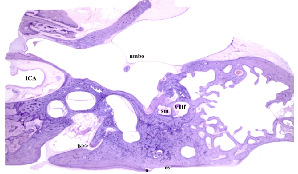
section281
The umbo of the tympanic membrane, which marks the inferior end of
the manubrium of the malleus, is visible. The stapedius muscle (sm) can be seen in the
posterior wall of the middle ear space. The endolymphatic sac (es) is present in the posterior
cranial fossa. The osseous opening of the canal, which transmits the endolymphatic duct, is
called the external aperture of the vestibular aqueduct. The medial lip of that aperture is
known as the “operculum” (o) (i.e., like the gill flap in fish). Nearly the entire course of the
foramen singulare (fs) is visible. The internal carotid artery (ICA) is seen in the carotid canal.
VIIf – Mastoid segment of facial nerve.
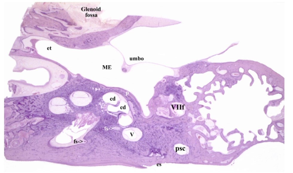
section 300
The hook portion of the cochlea duct (cd) is visible on the medial wall of the middle ear space. The promontory (p) which bulges into the middle ear space (ME) marks the position of the hook. Only a small part of the vestibule (v) remains. The end of the foramen singulare (fs) is visible. The non-sensory portion of the posterior semicircular canal (psc) is still present. The endolymphatic sac (es) is located in the posterior cranial fossa. et – Eustachian tube; VIIf – Mastoid segment of facial nerve.