岩尖胆脂瘤分型
Sanna 岩尖胆脂瘤分型
| Class | Location | Spread |
|
Class I: supralabyrinthine 迷路上型 |
geniculate ganglion of facial nerve |
anterior: horizontal part of ICA posterior: posterior bony labyrinth medial: IAC, petrous apex inferior: basal turn of the cochlea |
|
Class II: infralabyrinthine 迷路下型 |
hypotympanic and infralabyrinthine cells |
anterior: ICA vertical part, petrous apex, clivus posterior: dura of the posterior cranial fossa and sigmoid sinus medial: IAC, lower clivus, occipital condyle inferior: jugular bulb, lower cranial nerves |
|
Class III: infralabyrinthine-apical 迷路下-岩尖型 |
infralabyrinthine compartment, ICA reaching up to petrous apex |
anterior: ICA vertical 8 horizontal parts posterior: posterior fossa through the retrofacial air cells medial: petrous apex, clivus, sphenoid sinus, rhinopharynx inferior: jugular bulb, lower cranial nerves |
|
Class IV: massive labyrinthine 广泛型 |
entire otic capsule | anterior: ICA vertical 8 horizontal parts posterior: posterior fossa dura and IAC medial: petrous apex, superior and mid clivus, sphenoid sinus inferior: infralabyrinthine compartment |
|
Class V: apical 岩尖型 |
petrous apex | anterior: Meckel’s cave area and may involve the V nerve posterior: IAC and posterior cranial fossa medial: superior or mid clivus, sphenoid sinus inferior: infralabyrinthine compartment |
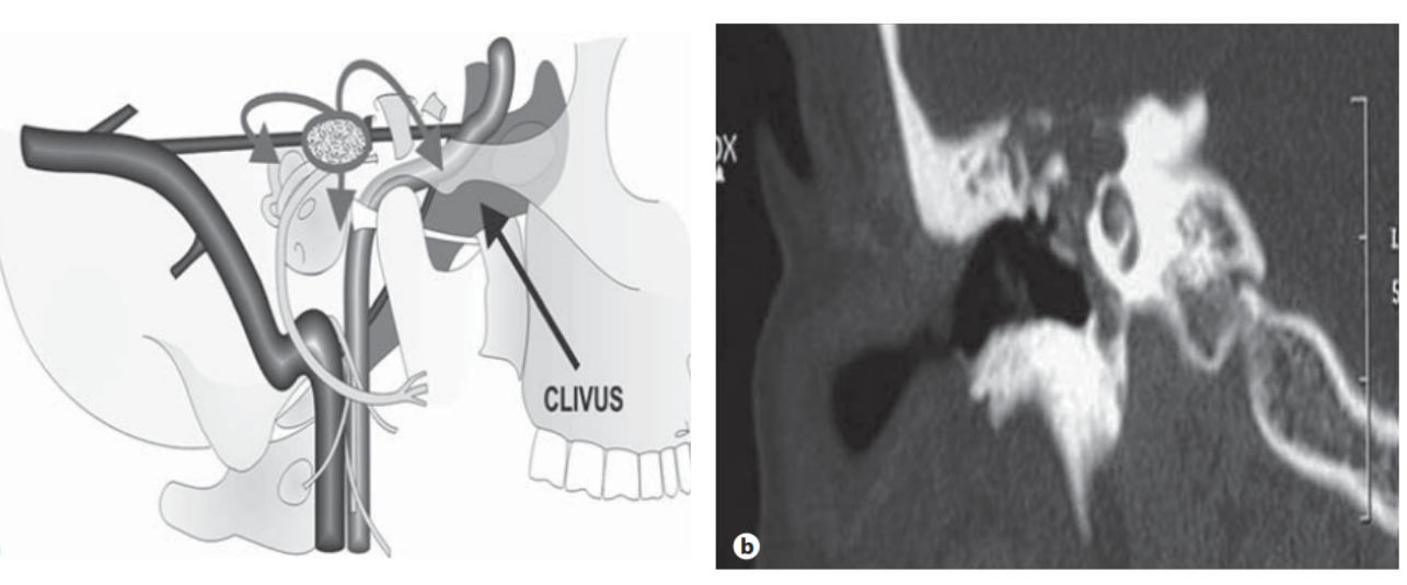
迷路上型
Fig. 1. a Diagrammatic representation of a supralabyrinthine PBC as viewed from the lateral aspect. The dotted ovoid area represents the site of the lesion and the arrows represent the route of spread. Directions for the route of spread are given in table 1. b Coronal HRCT image of the temporal bone showing a supralabyrinthine PBC. The dural plate is eroded. The cochlea is intact.

Fig. 2. a Diagrammatic representation of an infralabyrinthine PBC as viewed from the lateral aspect. The dotted area represents the site of the lesion and the arrows represent the route of spread. Directions for the route of spread are given in table 1. b Coronal HRCT image of the temporal bone showing an infralabyrinthine PBC.

Fig. 3. a Diagrammatic representation of an infralabyrinthine- apical PBC as viewed from the lateral aspect. The dotted area rep- resents the site of the lesion and the arrow represents the route of spread. Directions for the route of spread are given in table 1. b Coronal HRCT image of the temporal bone showing an infra- labyrinthine-apical PBC. Cholesteatoma involves the inferior as- pect of the internal auditory canal.
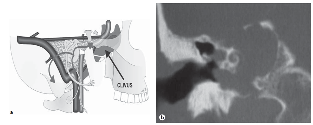
Fig. 4. a Diagrammatic representation of a massive PBC as viewed from the lateral aspect. The dotted area represents the site of the lesion and the arrows represent the route of spread. Directions for the route of spread are given in table 1. b Coronal HRCT image of the temporal bone showing a massive PBC. The cholesteatoma has eroded the cochlea extending into the petrous apex reaching up to the clivus. Cholesteatoma is eroding the dural plate.
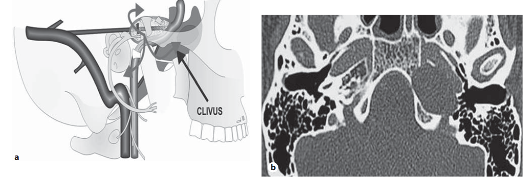
Fig. 5. a Diagrammatic representation of an apical PBC as viewed from the lateral aspect. The dotted area represents the site of the lesion and the arrows represent the route of spread. Directions for the route of spread are given in table 1. b An axial HRCT image of the temporal bone showing an apical PBC. There is a smooth osteolytic lesion occupying the petrous apex reaching up to the clivus, lying medial to the horizontal portion of the internal ca- rotid artery. Note that the bony canal of the horizontal portion of the internal carotid artery is eroded.
亚型
| Subclass | Features |
| Clivus (C) 斜坡亚型 | superior and mid clival extensions are seen from massive, infralabyrinthine-apical and apical PBC whereas the lower clival involvement is a feature of infralabyrinthine-apical PBC |
| Sphenoid sinus (S) 蝶窦亚型 | sphenoid sinus involvement is seen from anteromedial extensions of massive, infralabyrinthine-apical and apical PBC; it is a rare extension |
| Rhinopharynx (R) 鼻咽亚型 | it is the rarest extension of the PBC; it is an extension of infralabyrinthine-apical or massive PBC,which may extend through the clivus beneath the sphenoid sinus into the rhinopharynx |
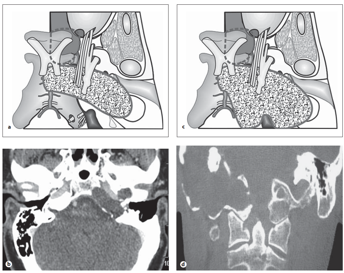
Fig. 6. a Diagrammatic representation of massive PBC with clival extension as seen from the superior aspect (see text). The choles- teatoma involves the petrous apex and superior and mid clivus. b Axial CT scan showing clival extension of a massive PBC. Note the radical mastoid cavity; this patient had previous surgery 15years prior to presentation. c Diagrammatic representation of ex- tension of massive PBC to clivus and occipital condyle as viewed from the superior aspect. d Coronal CT scan showing extension of massive PBC to clivus and occipital condyle.
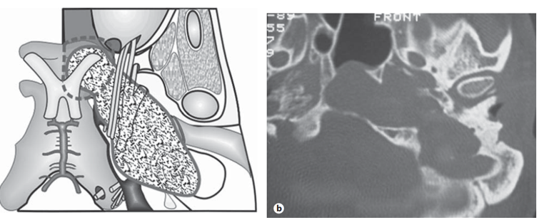
Fig. 7. a Diagrammatic representation of an infralabyrinthine- apical PBC with extension to the sphenoid sinus as viewed from the superior aspect. b Axial CT scan showing an infralabyrin- thine-apical PBC with extension to the sphenoid sinus.

Fig. 8. a Diagrammatic representation of a massive PBC with ex- tension to the rhinopharynx. The cholesteatoma passes beneath the sphenoid sinus to reach the rhinopharynx (see text). b Axial CT scan showing a massive PBC with extension to the rhinophar- ynx. c Coronal T 1 -weighted MRI showing extension of PBC into the rhinopharynx.